Home
Uncategories
Draw A Diagram Of Animal Cell And Label Centriole And Mitochondria On It : Diagram Of Animal Cell Anatomy Illustration Royalty Free Cliparts Vectors And Stock Illustration Image 103619517
Draw A Diagram Of Animal Cell And Label Centriole And Mitochondria On It : Diagram Of Animal Cell Anatomy Illustration Royalty Free Cliparts Vectors And Stock Illustration Image 103619517
Draw A Diagram Of Animal Cell And Label Centriole And Mitochondria On It : Diagram Of Animal Cell Anatomy Illustration Royalty Free Cliparts Vectors And Stock Illustration Image 103619517. Structure of a typical animal cell click to enlarge. Draw a squiggly line to show the folded inner membrane of the mitochondrian. Eukaryotic cells are larger, more complex, and have evolved more recently than prokaryotes. The animal cell diagram is widely asked in class 10 and 12 examinations and is beneficial to understand the structure and functions of an animal. Let's draw an animal cell:
Yr7 plant cell to label labelled diagram. Benda (1897) was the first to coin the term mitochondrion. The mitochondria are the powerhouses of the cell. Depending on your grade level you may add or remove some structures. Stain an onion cell (plant) using iodine and identify & draw a nucleus 5.
Plant Cell Definition Labeled Diagram Structure Parts Organelles from microbenotes.com Mitochondria, nucleus, nucleolus, golgi body, ser, rer, plasma membrane, centriole, ribosome, cytoplasm, answered by mhairi m. Draw an animal cell and label it draw a plant cell and label it draw a simple circle or oval for the cell membrane. Structure of a typical animal cell click to enlarge. There are three microtubules in each group. Draw and label a diagram of an animal cell? Draw a squiggly line to show the folded inner membrane of the mitochondrian. Within a cell you have the cell membrane which is the outer wall of the cell, you also have the nucleolus, nucleus, golgi body, vacuole, lysosome and other parts to the cell that you will see pointed poit for you in the first step of this tut. Plant and animal cells ks3 revision.
Yes plants have a cell wall and a cell membrane but animals dont have a cell wall they only have a cell membrane asked in human anatomy and physiology draw structure of human cell and label.
Label and show the locations of the following organelles on the diagram of an animal cell below: Draw a squiggly line to show the folded inner membrane of the mitochondrian. The outer membrane and the inner membrane are made of proteins and phospholipid layers. From amoebae to earthworms to mushrooms, grass. Yr7 plant cell to label labelled diagram. Animal cells contain organelles known as centrioles which are not present in plant cells. The animal cell diagram is widely asked in class 10 and 12 examinations and is beneficial to understand the structure and functions of an animal. Microtubules (and centrioles) are part of the cytoskeleton. On the contrary plant cells lack centrioles and intermediate filaments which are present in animal cells. Draw a diagram of an animal cell and label at least eight organelles in it. It helps in carrying out the functions such as respiration, nutrition, digestion, excretion etc. All cells are made up of protoplasm, which consists of water, electrolytes, proteins, lipids, and carbohydrates. Animals don't rely on this water storage for the rigidity of their form, and use their vacuoles.
Its size ranges from 0.5 to 1.0 micrometre in diameter. It is the nucleus mitochondrion chloroplast golgi apparatus 6. Use the zoom slider to see the cell at a magnification of 1000x (1000 times larger than normal). Mitochondria are present in plant and animal cells. Draw and label an animal cell.
1 Draw A Neat Labelled Diagram Of Animal Cell And List The Function Of Each Organalle Brainly In from hi-static.z-dn.net Use the up/down and left/right sliders to manipulate the cell. Calculate the model sizes and draw and label a scaled diagram of the organelles of the cell that will fit on another sheet of graph paper and can be drawn with some detail (i.e. Mitochondria are commonly called the power house of the cell. A cell model is the size of this classroom (approximately 10 m in length). Draw an animal cell and label it draw a plant cell and label it draw a simple circle or oval for the cell membrane. Animals don't rely on this water storage for the rigidity of their form, and use their vacuoles. Each vacuole is separated from the cytoplasm by a single unit membrane, called the tonoplast. Where, prokaryotes are just bacteria and archaea, eukaryotes are literally everything else.
On the contrary plant cells lack centrioles and intermediate filaments which are present in animal cells.
Mitochondria are present in plant and animal cells. One of the distinctive aspects of a plant cell is the presence of a cell wall outside the cell membrane. Each vacuole is separated from the cytoplasm by a single unit membrane, called the tonoplast. So it is called as the structural and functional unit of life. Plant cell and animal cell diagram. A simple diagram of a plant leaf cell labelled in english. Structure of a typical animal cell click to enlarge. In the complete animal cell centrosome, the two centrioles are arranged such that one is perpendicular to. Examining a diagram of the plant cell will help make the differences clearer. Label the animal cell diagram, with a glossary of animal cell terms on a separate page. 1.4.1 draw a diagram to show the fluid mosaic model of a biological membrane note: Those are the main parts of a cell in an animal that you will have to draw. Label and show the locations of the following organelles on the diagram of an animal cell below:
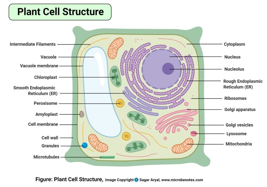
A plant cell diagram like the one above shows each part of the plant cell including the chloroplast cell wall plasma membrane nucleus mitochondria ribosomes etc. Label and show the locations of the following organelles on the diagram of an animal cell below: Use the zoom slider to see the cell at a magnification of 1000x (1000 times larger than normal). To start, click sample to take a sample of an animal cell. Investigate and draw an onion cell (plant) and a cork cell.
Label And Color The Animal And Plant Cells Pdf Cell Nucleus Endoplasmic Reticulum from imgv2-1-f.scribdassets.com Mitochondria are present in plant and animal cells. 2.3 eukaryotic cellsedit 2.3.1 draw and label a diagram of the ultrastructure of a liver cell as an example of an animal cell diagram of an animal cell should include free ribosomes, the rough endoplasmic reticulum (rer), lysosome, golgi apparatus, mitochondrion, and nucleus. Draw and label an animal cell. Each mitochondrion (singular) should contain an enclosed shape with many ridges and switchback lines. Eukaryotic cells are larger, more complex, and have evolved more recently than prokaryotes. Draw the animal cell and label the parts drawing an animal cell. Diagram your mitochondria like beans with a cross section showing the folds of the inner membrane. Use the up/down and left/right sliders to manipulate the cell.
Pick a representative object that is commonly found and has the same size as that of your
Depending on your grade level you may add or remove some structures. One of the distinctive aspects of a plant cell is the presence of a cell wall outside the cell membrane. Structures unique to animal cells. Benda (1897) was the first to coin the term mitochondrion. Draw an animal cell and label it draw a plant cell and label it draw a simple circle or oval for the cell membrane. Pick a representative object that is commonly found and has the same size as that of your Eukaryotic cells are larger, more complex, and have evolved more recently than prokaryotes. * mitochondria supply the energy for the majority of cells. Stain an onion cell (plant) using iodine and identify & draw a nucleus 5. It is the nucleus mitochondrion chloroplast golgi apparatus 6. 1.4.1 draw a diagram to show the fluid mosaic model of a biological membrane note: The unit of life > eukaryotic cells > draw a diagram of an animal. Animals don't rely on this water storage for the rigidity of their form, and use their vacuoles.
Structures unique to animal cells draw a diagram of animal cell. The cell structure gizmo™ allows you to look at typical animal and plant cells under a microscope.


0 Comments:
Posting Komentar