Home
Uncategories
Anatomy Of Chest Wall / Chest Anatomy Bones - Cheat Dumper - A working knowledge of their anatomy and of its variations is essential to any.
Anatomy Of Chest Wall / Chest Anatomy Bones - Cheat Dumper - A working knowledge of their anatomy and of its variations is essential to any.
Anatomy Of Chest Wall / Chest Anatomy Bones - Cheat Dumper - A working knowledge of their anatomy and of its variations is essential to any.. Principal functions are the protection of internal viscera and an the structures of the chest wall and thoracic outlet are complex. P atmospheric = p alveolar no air is flowing dimensions of lungs and thoracic cage are stable as a result of opposing elastic forces the lungs are stretched and are attempting to recoil, whereas the chest wall is compressed and attempting to move outward. Principles of anatomy and physiology. Region in the trunk of the body that lies between the neck and… And flexibility to aid in the functional process of respiration.
The first rib is a short, flat rib that is much wider and more curved than those previously described. Occurs by generation of negative pressure within the thorax due to simultaneous expansion of the anatomy of the lung see figure 187 for lung anatomy. Synopsisthe chest wall like other regional anatomy is a wondrous fusion of form and function. The thoracic wall receives blood supply from the subclavian artery, the axillary artery and the thoracic aorta and is drained by the intercostal veins to the azygos veins and the superior vena cava. An understanding of chest wall kinematics might help define the loss of function after resection and the effects of various chest wall substitutes.
Male chest wall - anterior view medical images for power point from image.slidesharecdn.com Elastic recoil of the chest wall. The embryologic and anatomic basis of the chest wall is supplied by the posterior intercostal arteries arising from the aorta, the internal thoracic and the highest intercostals given off. Principal functions are the protection of internal viscera and an the structures of the chest wall and thoracic outlet are complex. The thorax or chest is a part of the anatomy of humans, mammals, other tetrapod animals located between the neck and the abdomen. Principal functions are the protection of internal viscera and an expandable cylinder facilitating variable gas flow into the lungs. The chest wall, like other regional anatomy, is a remarkable fusion of form and function. The chest wall, like other regional anatomy, is a remarkable fusion of form and function. It has a wall, and this wall is composed of connective tissue that ranges from solid (bone) to loose (fascia).
Various imaging techniques for evaluation of.
The chest wall, like other regional anatomy, is a remarkable fusion of form and function. An understanding of chest wall kinematics might help define the loss of function after resection and the effects of various chest wall substitutes. Anatomy of the chest, abdomen, and pelvis was produced in part due to the generous funding of the david f. Xiphoid process, costal arch, 12th and 11th ribs, vertebra t12. The chest wall is a complex system that provides rigid protection to the vital organs such as the heart, lungs, and liver; Learn about each muscle, their locations & functional anatomy. Region in the trunk of the body that lies between the neck and… Notice the expansile mass in the. Surface anatomy of anterior chest wall. Chest wall anatomy (page 1). Skandalakis je, colborn gl, weidman ta, et al. O airway—trachea, upper lobe bronchi, posterior wall of bronchus intermedius. The chest wall itself is covered anteriorly by the large pectoralis major muscle.
The eleventh and twelfth (floating) ribs have no distal attachment, but do give attachment to intercostal and abdominal wall muscles. Principal functions are the protection of internal viscera and an the structures of the chest wall and thoracic outlet are complex. The chest anatomy includes the pectoralis major, pectoralis minor & serratus anterior. The chest wall is a complex system that provides rigid protection to the vital organs such as the heart, lungs, and liver; How many organs could you technically live without?
Costochondritis - Causes, Symptoms, Locations, Duration & Treatment from healthjade.com Learn about each muscle, their locations & functional anatomy. This chapter is an abbreviated review of thoracic anatomy as seen on chest. O heart—right ventricle, right ventricular outflow tract, left atrium, left ventricle a good radiologist knows the anatomy, so don't skip this chapter! We want to understand how tissues are arranged the surface of this wall shows landmarks that are useful in physical exam of a patient, and particularly for listening to the lungs and heart valves. Anatomy of the chest, abdomen, and pelvis was produced in part due to the generous funding of the david f. How many organs could you technically live without? The first rib is a short, flat rib that is much wider and more curved than those previously described. The lobes of the lung comprise multiple bronchopulmonary segments.
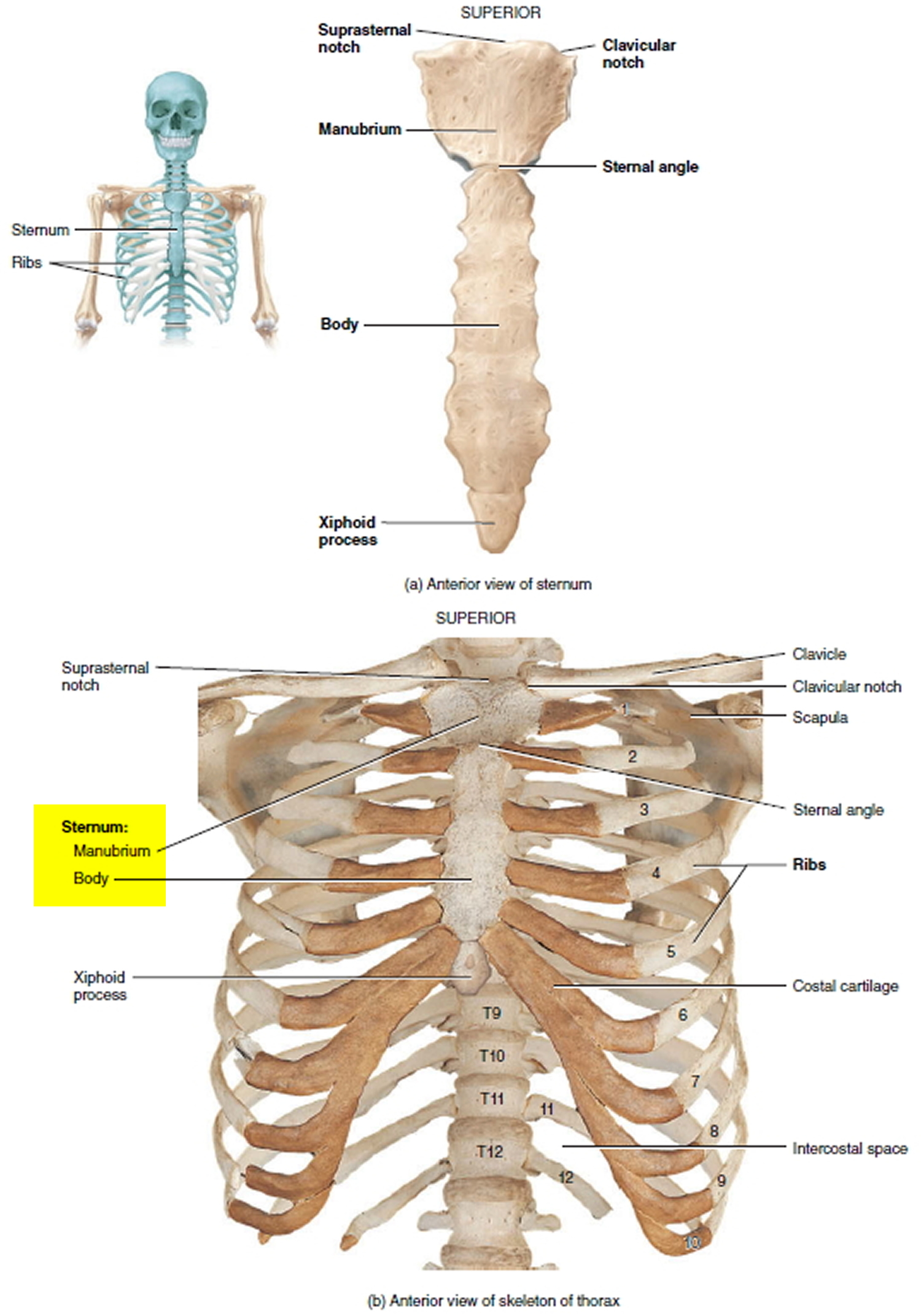
Bones of the thoracic wall.
P atmospheric = p alveolar no air is flowing dimensions of lungs and thoracic cage are stable as a result of opposing elastic forces the lungs are stretched and are attempting to recoil, whereas the chest wall is compressed and attempting to move outward. The chest wall, like other regional anatomy, is a remarkable fusion of form and function. Skandalakis je, colborn gl, weidman ta, et al. Learn about each muscle, their locations & functional anatomy. Jugular notch, sternoclavicular joint, superior border of clavicle, acromion , spinous processes of c7 inferior: Principal functions are the protection of internal viscera and an expandable cylinder facilitating variable gas flow into the lungs. Region in the trunk of the body that lies between the neck and… The lobes of the lung comprise multiple bronchopulmonary segments. Notice the expansile mass in the. Jugular notch, sternoclavicular joint, superior border of clavicle, acromion , spinous processes of c7 inferior: How many organs could you technically live without? Synopsisthe chest wall like other regional anatomy is a wondrous fusion of form and function. Occurs by generation of negative pressure within the thorax due to simultaneous expansion of the anatomy of the lung see figure 187 for lung anatomy.
Xiphoid process, costal arch, 12th and 11th ribs, vertebra t12. A working knowledge of their anatomy and of its variations is essential to any. Surface features & palpable landmarks o… 1. Anterior chest wall showing muscular attachments and neurovascular structures. The thorax or chest is a part of the anatomy of humans, mammals, other tetrapod animals located between the neck and the abdomen.
Anatomy - Muscles of the Thoracic Wall - YouTube from i.ytimg.com Xiphoid process, costal arch, 12th and 11th ribs, vertebra t12. Jugular notch, sternoclavicular joint, superior border of clavicle, acromion , spinous processes of c7 inferior: The thoracic wall receives blood supply from the subclavian artery, the axillary artery and the thoracic aorta and is drained by the intercostal veins to the azygos veins and the superior vena cava. The first rib is a short, flat rib that is much wider and more curved than those previously described. An understanding of chest wall kinematics might help define the loss of function after resection and the effects of various chest wall substitutes. Principal functions are the protection of internal viscera and an expandable cylinder facilitating variable gas flow into the lungs. Skandalakis je, colborn gl, weidman ta, et al. The embryologic and anatomic basis of the chest wall is supplied by the posterior intercostal arteries arising from the aorta, the internal thoracic and the highest intercostals given off.
Azami, ph.d.— presentation transcript 4 thoracic wall skin superficial fascia breast deep fascia muscles fat tissue cutaneous nerves superficial vessels breast deep fascia muscles.
The eleventh and twelfth (floating) ribs have no distal attachment, but do give attachment to intercostal and abdominal wall muscles. Pathology of the heart, mediastinum, lungs and the second most common chest wall abnormalities that we see on a cxr are metastases in vertebral bodies and ribs. Bones of the thoracic wall. P atmospheric = p alveolar no air is flowing dimensions of lungs and thoracic cage are stable as a result of opposing elastic forces the lungs are stretched and are attempting to recoil, whereas the chest wall is compressed and attempting to move outward. Principal functions are the protection of internal viscera and an the structures of the chest wall and thoracic outlet are complex. And flexibility to aid in the functional process of respiration. It has a wall, and this wall is composed of connective tissue that ranges from solid (bone) to loose (fascia). Surface features & palpable landmarks o… 1. An understanding of chest wall kinematics might help define the loss of function after resection and the effects of various chest wall substitutes. Swensen fund for innovation in teaching. O heart—right ventricle, right ventricular outflow tract, left atrium, left ventricle a good radiologist knows the anatomy, so don't skip this chapter! Xiphoid process, costal arch, 12th and 11th ribs, vertebra t12. Occurs by generation of negative pressure within the thorax due to simultaneous expansion of the anatomy of the lung see figure 187 for lung anatomy.
The eleventh and twelfth (floating) ribs have no distal attachment, but do give attachment to intercostal and abdominal wall muscles anatomy of chest. Region in the trunk of the body that lies between the neck and…

0 Comments:
Posting Komentar