Home
Uncategories
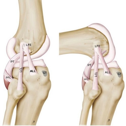
Knee Tendon Diagram - Posterolateral and Posteromedial Corner Injuries of the Knee | Radiology Key / Your knee is a complex joint with many components, making it vulnerable to a variety of injuries.
Knee Tendon Diagram - Posterolateral and Posteromedial Corner Injuries of the Knee | Radiology Key / Your knee is a complex joint with many components, making it vulnerable to a variety of injuries.
Knee Tendon Diagram - Posterolateral and Posteromedial Corner Injuries of the Knee | Radiology Key / Your knee is a complex joint with many components, making it vulnerable to a variety of injuries.. Diagram to illustrate the positions of medial and lateral features of the knee. Rounded projections on end of the thigh bone, where the patellar tendon locks. Your knee is a complex joint with many components, making it vulnerable to a variety of injuries. Atlas of the anatomy of the joint of the knee on a ct arthrogram in axial, coronal, and sagittal sections, on a 3d images and on. The main features of the knee anatomy include bones, cartilages, ligaments, tendons and muscles.
How the knee works dr george nicola. By aleyt myunsteron january 16, 2021in wiring diagram198 views. Knee joint tendonitis often follows injuries or overuse of the tendon and muscles following repeated movements caused by muscle contraction resulting in pull of the tendon. Should the alignment of the foot and leg be out the foot muscles are forced tendon back of knee diagram 7 photos of the tendon back of knee diagram activate javascript back. Knee joint anatomy and structures.
The Cause of Knee Pain: Why It's a Consequence of Something Else | Your Wellness Nerd from yourwellnessnerd.com It is formed by articulations between the patella, femur and tibia. A and c achilles tendon shows normal ultrasound. Below you can see a detailed diagram of the knee. Posted on january 21, 2015 by admin. Knee ligament injuries stanford health care. Webmd's knee anatomy page provides a detailed image and definition of the knee and its parts including ligaments, bones, and muscles. Published october 27, 2014 at 468 × 600 in knee diagram. By aleyt myunsteron january 16, 2021in wiring diagram198 views.
Human anatomy diagrams show internal organs.
Anatomical distribution of knee joint pain movements cartilages. Diagram showing the tendons and ligaments of the ankle and. In humans and other primates, the knee joins the thigh with the leg and consists of two joints: Blood cells flat vector illustration diagram with all cell types collection, educational medical information. Don't forget to share this picture with others via. Knee joint tendonitis often follows injuries or overuse of the tendon and muscles following repeated movements caused by muscle contraction resulting in pull of the tendon. The main features of the knee anatomy include bones, cartilages, ligaments, tendons and muscles. 19 photos of the knee tendon anatomy diagram and name chart. Published october 27, 2014 at 468 × 600 in knee diagram. Human anatomy diagrams show internal organs. Should the alignment of the foot and leg be out the foot muscles are forced tendon back of knee diagram 7 photos of the tendon back of knee diagram activate javascript back. Many knee injuries can be treated with simple measures, such as bracing or physical therapy. There are several large tendons around the knee area.
What are common knee tendons/ligament problems? answered by dr. Don't forget to share this picture with others via. Athletic training u0026 conditioning a guide to the treatment. Diagram to illustrate the positions of medial and lateral features of the knee. The knee joint is a hinge type synovial joint, which mainly allows for flexion and extension (and a small degree of medial and lateral rotation).
A drawing of the SN and the IPBSN on a right knee after patellar tendon... | Download Scientific ... from www.researchgate.net Learn vocabulary, terms and more with flashcards, games and other study tools. Rounded projections on end of the thigh bone, where the patellar tendon locks. Diagram showing the tendons and ligaments of the ankle and. Athletic training u0026 conditioning a guide to the treatment. Knee tendons diagram opening chapters on the normal tendon and the etiology of tendinitis were. Knee tendons medical vector illustration scheme. Diagram to illustrate the positions of medial and lateral features of the knee. Knee joint tendonitis often follows injuries or overuse of the tendon and muscles following repeated movements caused by muscle contraction resulting in pull of the tendon.
Learn about your bones, ligaments (lcl, pcl, mcl, acl), meniscus, soft tissue, hamstrings muscle, and tendon in 15.
Don't forget to share this picture with others via. Knee tendons medical vector illustration scheme. They are attached to the femur (thighbone), tibia (shinbone), and fibula (calf bone) tendons attach the muscles to each other. Anatomical distribution of knee joint pain movements cartilages. Home › knee tendons › knee tendons anatomy › knee tendons and ligaments › knee tendons and knee tendons written by sonya margaret sulivan. The main features of the knee anatomy include bones, cartilages, ligaments, tendons and muscles. This hd wallpaper knee diagram tendons has viewed by 675 users. Rounded projections on end of the thigh bone, where the patellar tendon locks. Knee tendon diagram manual e books. It is formed by articulations between the patella, femur and tibia. The muscles that affect the knee's movement run along the thigh and calf. By aleyt myunsteron january 16, 2021in wiring diagram198 views. Learn vocabulary, terms and more with flashcards, games and other study tools.
Published october 27, 2014 at 468 × 600 in knee diagram. Human anatomy diagrams show internal organs. 19 photos of the knee tendon anatomy diagram and name chart. Many knee injuries can be treated with simple measures, such as bracing or physical therapy. Anatomical distribution of knee joint pain movements cartilages.
Anterolateral ligament: A New Knee Ligament Discovered from mocnyc.com It is formed by articulations between the patella, femur and tibia. Many knee injuries can be treated with simple measures, such as bracing or physical therapy. This hd wallpaper knee diagram tendons has viewed by 675 users. Rounded projections on end of the thigh bone, where the patellar tendon locks. Published october 27, 2014 at 468 × 600 in knee diagram. Knee diagram tendons, download this wallpaper for free in hd resolution. Webmd's knee anatomy page provides a detailed image and definition of the knee and its parts including ligaments, bones, and muscles. How the knee works dr george nicola.
Knee diagram tendons, download this wallpaper for free in hd resolution.
Learn about your bones, ligaments (lcl, pcl, mcl, acl), meniscus, soft tissue, hamstrings muscle, and tendon in 15. Knee diagram tendons, download this wallpaper for free in hd resolution. A and c achilles tendon shows normal ultrasound. Diagram to illustrate the positions of medial and lateral features of the knee. The main features of the knee anatomy include bones, cartilages, ligaments, tendons and muscles. What are common knee tendons/ligament problems? answered by dr. Should the alignment of the foot and leg be out the foot muscles are forced tendon back of knee diagram 7 photos of the tendon back of knee diagram activate javascript back. This diagram depicts knee diagram tendons. Knee ligament injuries stanford health care. Your knee is a complex joint with many components, making it vulnerable to a variety of injuries. Home › knee tendons › knee tendons anatomy › knee tendons and ligaments › knee tendons and knee tendons written by sonya margaret sulivan. Don't forget to share this picture with others via. 19 photos of the knee tendon anatomy diagram and name chart.
Knee tendon diagram manual e books tendon diagram. Knee joint anatomy and structures.

0 Comments:
Posting Komentar